Various imaging methods are used to capture information about tissue, cell and subcellular structures. However, a single method can only capture information about tissue structure or function, and detailed observation on the nano scale means that scientists lose information about the wider environment. In other words, in order to gain a comprehensive understanding of tissue, scientists need to combine imaging technology.
In the new study, scientists developed a method that combines seven imaging methods, including in vivo imaging, synchrotron radiation X-ray and volume electron microscopy. They demonstrated their method by imaging two different regions of the mouse brain - the olfactory bulb and the hippocampus.
Importantly, the technology can be applied to other regions of the brain or other parts of the body and provide scientists with a more detailed understanding of many different biological structures and tissues.
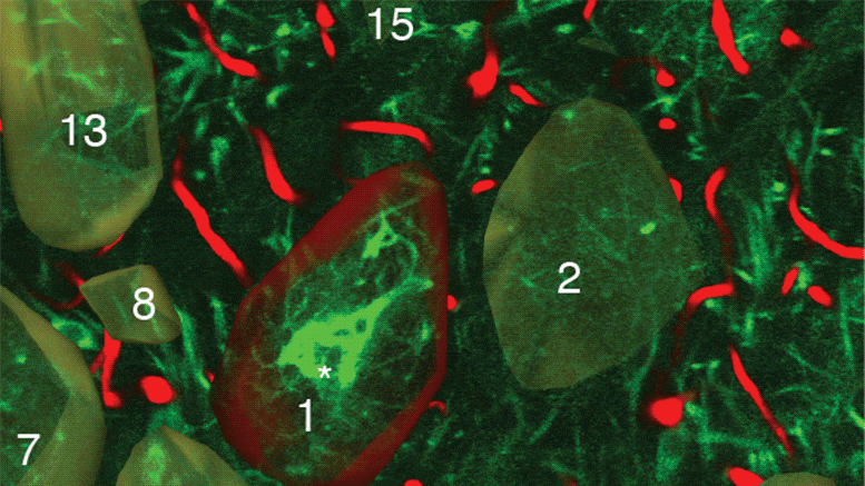
Each step of the imaging process provides different information. First, the researchers used in vivo calcium imaging to visualize neurons in specific areas of the brain and see which neurons were active when the mice were exposed to odor.
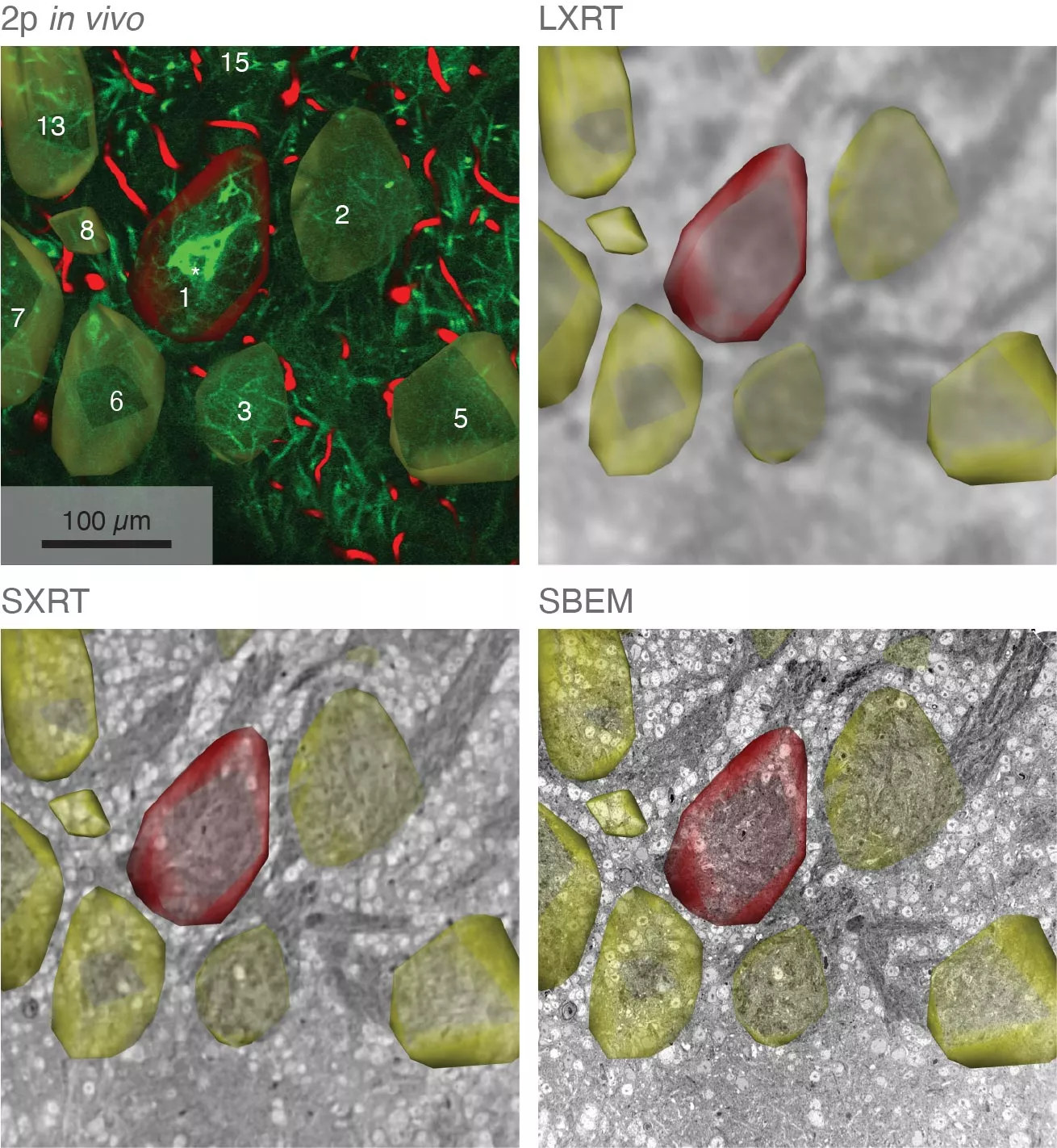
After the mice were euthanized, scientists used various methods to image brain tissue samples, including synchrotron radiation X-ray tomography, which can capture samples up to several millimeters long. This scale is large enough for scientists to see the entire neural network and the location of specific cells or other structures in a wider range of samples. It is important that this method does not damage the sample, so it can be imaged again with another technique.
The researchers then selected areas of particular interest and imaged them with an electron microscope to capture complex details at high resolution. In some target areas, this can map details as small as 10 nanometers, allowing researchers to see tiny structures, such as a single synapse connecting neurons.
Using computer algorithms, the researchers combined these results and drew a complete map of the structure and function of the parts of the brain they studied. It is reported that the maximum can reach a few cubic millimeters.
Carles Bosch, the first author of the research paper from the laboratory of sensory circuits and neural technology at Crick University and the chief laboratory research scientist, pointed out: "our method provides a reliable method to overcome the challenge of imaging structures on huge and different scales. We believe it will become a powerful tool to study the structure and function of neuronal circuits and other tissues in the mammalian brain."
Senior author of Crick University Andreas Schaefer, head of the laboratory of sensory circuits and neurotechnology, said: "We are very interested in applying this method to the brain, where, in addition to the information about specific neurons and synapses, it is also very important to collect information about the entire neural network, which is a few millimeters long. But it also has great potential to play a role in other environments, such as cancer biology, and the purpose of researchers is to understand the activities of specific cells in a broader tumor context."
It is reported that the research team will continue the research and use this imaging method to find more details about the olfactory bulb. Of course, they will make further efforts to improve the technology.